
Test of Practical on Stem Anatomy of Dicotyledoneae and Monocotyledoneae Plants.
Specimen of Study.
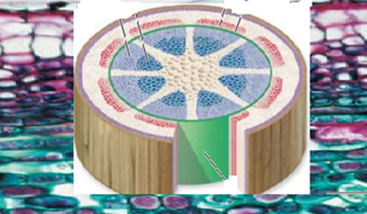
- TS of a permanent slide of a dicot stem, such as a Sida acuta
- TS of a temporary slide of the stem of Tridax mounted in water or dilute glycerin under the microscope
- TS of Tridax mounted in iodine solution or any suitable stain under the microscope
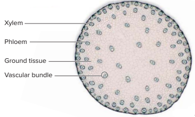
- TS of maize plant mounted in iodine solution or any suitable stain under the microscope
Note, however, which tissues stain which colour.
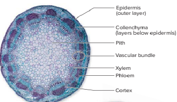
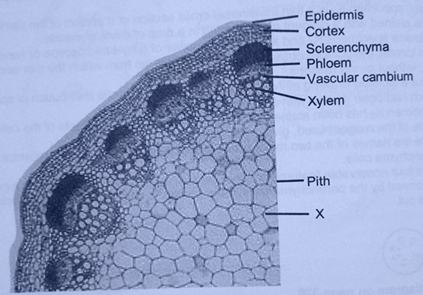
The primary tissues in the dicotyledonous stem are;
- Epidermis
- Cortex
- Vascular cylinder
- Pith
The Epidermis
- It is the outermost layer around the stem.
- It is a single layer of closely packed cells
- The cells lack chloroplasts
- The outer wall of the cells is thickened with a waxy cuticle.
- Some epidermal cells may bear epidermal hairs.
The cortex
- The cortex is found beneath the epidermis.
- It consists of three types of cells (tissues).
- An outer collenchyma, middle parenchyma, and inner endodermis.
- The outer layer is made up of collenchyma cells.
- The cells are small, tightly packed cells, thickened at the corners.
- The second layer is made up of parenchyma cells.
- These cells are large, thin-walled, with narrow air spaces between them.
- The innermost layer is the endodermis
- It is made up of a single layer of rectangular, closely fitting cells
- It forms a ring around the vascular tissue
- The cells contain starch grains and make the endodermis stain blue-black with iodine solution.
It should be noted that:
- The endodermis is less visible in the stem than in the root.
- The endodermis is referred to as the starch sheath.
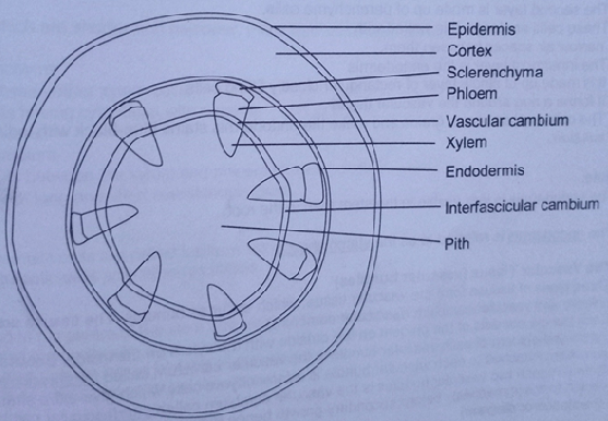
The Vascular Tissue (Vascular Bundles/Vascular Cylinder)
- Three types of tissues form the vascular tissue, which occur as bundles.
- The tissues are: xylem, phloem and vascular cambium (fascicular cambium)
- Each bundle consists of the phloem on the outside with the xylem on the inside.
- Between the xylem and phloem of each vascular bundle is the vascular cambium, called the intrafascicular cambium.
- Attached to each vascular bundle are sclerenchyma cells, which give extra strength.
- Between each two vascular bundles is the vascular cambium, called the interfascicular cambium (which forms immediately before secondary growth, hence may not be necessary in such description or diagram).
The phloem
The phloem consists of three types of cells, namely;
- sieve tubes,
- companion cells and
- phloem parenchyma.
- Sieve tubes are elongated cells that live and have cytoplasm but no nucleus at maturity.
- The cells are arranged end to end with perforated cross-walls called sieve plates.
- Beside each sieve tube is the companion cell.
- Companion cells are narrow cells having dense cytoplasm and a nucleus.
The xylem
This is made up of the
- Vessel elements,
- tracheid,
- fibres and
- parenchyma
Vessel Elements
- The vessel elements are the main conducting tubes of the xylem.
- The cells are dead at maturity.
- They are elongated tubes with no cross-walls.
- The cell walls are lignified.
Tracheid
- Tracheids are dead cells at functional maturity
- They are elongated with a tapering end.
- Cell walls lignified.
- The cell walls have pits and depressions.
Fibres
- Dead cells, which are shorter and narrower, more lignified/thicker cell wall.
Xylem Parenchyma
- It occurs between other xylem cells.
- Living cells have a cytoplasm with a nucleus.
- Thin cell walls.
Vascular Cambium
- This occurs between the xylem and phloem of each bundle.
- It consists of long and short meristematic cells.
Pith
- This is found at the centre of the stem and extends between the bundles.
- It consists of thin-walled parenchyma cells with air spaces.
- Medullary rays provide lateral transfer of materials between the vascular bundles and the inner(pith) and outer parts(cortex) of the stem
It should be noted that;
- Long meristematic cells are called fusiform initials
- Short meristematic cells are called ray initials
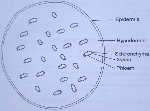
Differences between monocotyledonous and dicotyledonous stems
| Monocotyledonous Stem | Dicotyledonous Stem |
| Presence of pith | Absence of pith. |
| Vascular bundles form a ring. | Vascular bundles are scattered in the ground tissue. |
| Presence of endodermis | Absence of endodermis. |
| Vascular cambium is present. | Vascular cambium absent |
| Presence of many xylem and phloem cells | Presence of a few xylems and phloem cells |
| Have a wide cortex. | Have a narrow cortex. |
STUDY QUESTIONS.
-
Study the diagram below and answer the questions that follow.
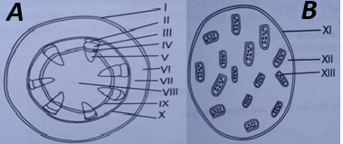 a. Differentiate between the two specimens.
b. Two examples each of plants from which the specimens can be obtained.
c. Identify four tissues in specimen A.
d. Give one function of each tissue named.
e. Give two functions common with specimens A and B.
f. Which tissue stains blue black with a laboratory reagent.
- Name the reagent
- Give one reason
h. Which tissue is for cell division.
i. Which two tissues provide support?
j. Give one common name for the parts labelled VI and XIII.
k. Name the cells in each of the parts VI, VII and XII.
2. Study the diagram below and answer the questions that follow
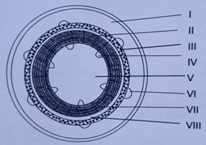
- Identify the labelled parts from I to VIII.
- State the function of VI and IV.
3. study carefully the specimen below as observed under the high power of microscope and answer the questions that follow.
a) Identify the specimen
b) Give two reasons for your answer
c) Name five tissues
d) Give one function of each tissue named
e) Give any two examples of organisms from which the specimen can
f) Name one stain used in the preparation of the specimen
g) Name the single cell represented as X in the specimen.